The Human Brain
 The part of the central nervous system enclosed by the skull. Like its continuation the spinal cord, the brain starts life as a simple tube. But whereas the spinal cord retains this form throughout life, the brain grows into a complex system of bulges and folds.
At an early- stage of embryonic life, three bulbous swellings appear at the front end of the tube that is to form the central nervous system, and these develop into the three main divisions of the brain: the hind brain, the mid brain, and the fore brain.
The original tube is still recognizable in the adult brain as the brain-stem, seen as an extension into the skull of the spinal cord. It is a central core from which the larger portions of the brain (cerebellum and cerebrum) grow.
The brain-stem is the direct upward extension of the spinal cord into the skull, composed of medulla oblongata, pons, and mid brain. The major portions of the brain, cerebellum and cerebral hemispheres, project from it.
Very roughly speaking, the further from the spinal cord the more advanced the function, progressing from reflexes for maintaining vital functions such as the circulation of blood and breathing in the lower part of the hindbrain to the processes of intelligent behaviour in the outer layers of the forebrain. But structure and function are only loosely related, for the whole nervous system works as an integrated unit.
1. HIND BRAIN
The spinal cord continues into the skull as the medulla oblongata. In the cord, the nerve cells form a fluted rod (grey matter) surrounded by nerve fibres (white matter). In the medulla the grey matter is gathered into more or less distinct groups of cells or nuclei; this tendency increases further up the brain. These nuclei include the cells of several cranial nerves and also the vital centres which regulate fundamental activities such as the heart beat.
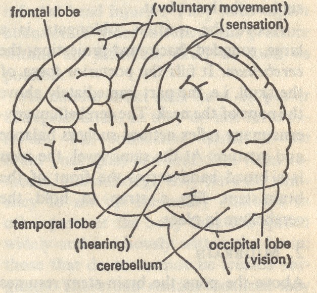
Above the medulla oblongata is a large, rounded, backward projection, the cerebellum. It fills the posterior fossa of the skull, i.e. the part immediately above the nape of the neck. The cerebellum governs many reflex actions such as balance and posture. At the same level, the pons is a broad band across the front of the brain-stem, like a strap to hold the cerebellum in place.
2. MID BRAIN
Above the pons the brain-stem resumes its primitive tubular structure. The midbrain is a short cylinder from which the stalks of the two cerebral hemispheres (forebrain) arise. It contains the cells of some cranial nerves, relay stations for the senses of sight and hearing, and centres for the coordination of movements. The reticular formation is a crisscross arrangement of grey and white matter. It can be traced through the whole length of the brain-stem but is most obvious in the centre of the midbrain. It appears to act as a central switchboard for the whole brain.
3. FORE BRAIN
Above the mid brain the brain-stem bends sharply forwards, and the narrow central canal widens to a deep vertical cleft, the third ventricle. At each side of the ventricle is a mass of grey matter, the thalamus. Sensation of all kinds is received here and distributed to reflex pathways or to the cerebral cortex and consciousness. Crude sensation, particularly pain, is perceived in the thalamus itself. Below is the hypothalamus, the highest centre of the autonomic nervous system, from which is suspended the pituitary gland. Behind is a small protrusion, the pineal body. The whole complex, representing the headward end of the brainstem, is sometimes called the tween brain.
Projecting from either side of the tween-brain and completely enclosing it are the relatively huge cerebral hemispheres, which make up the greater part of the human brain. It is only through the development of the cerebral hemispheres that a man's brain is relatively larger than a monkey's or a monkey's than a dog's. (An elephant's brain is about four times as large as man's, but in relation to the size of the animal it is much smaller.).
The central cavity of a cerebral hemisphere, the lateral ventricle, opens by a narrow isthmus (foramen of Monro) into the third ventricle. It is surrounded by masses of grey matter, the basal ganglia, which are concerned with muscle action immediately below the level of consciousness. Disorders of the basal ganglia cause parkinsonism - shaking palsy. The surface of the hemispheres is covered with a layer of grey matter, the cerebral cortex. Between the cortex and the underlying basal ganglia and thalamus is a broad zone of white matter, composed of nerve fibres to and from the cells of the cortex.
Definite functions can be ascribed to certain areas of the cerebral cortex. The areas for voluntary movement and for most types of sensation have been plotted in detail, but no particular area can be allotted to such functions as reason or memory.
4. SUPPORTING STRUCTURES
Nervous tissue needs a large and constant supply of oxygen and glucose, whether waking or sleeping, thinking or idling. It ceases to work after a few seconds if the supply is cut off, and irreparable damage is done in a few minutes. The rate of circulation in other organs varies widely but the brain receives about 750 nil of blood per minute, by the carotid and vertebral arteries, regardless of what is going on elsewhere. The complicated mechanisms for maintaining the blood pressure serve mainly to keep up this supply.
The brain is enclosed by fibrous membranes, the meninges, and by the cerebro spinal fluid.
The behaviour of nervous tissue, of which the brain is composed, is discussed under nervous system.
5. DISORDERS
Numerous kinds of defective or faulty development account for most of the severe cases of congenital mental handicap. A relatively small defect sometimes obstructs the circulation of the cerebro spinal fluid through and around the brain, causing a dangerous accumulation of fluid under pressure within the skull (hydrocephalus). This can often be corrected surgically.
Injuries to the brain are never to be taken lightly The mildest recognizable injury is concussion, and even the briefest loss of consciousness represents some permanent damage. Brain cells, once damaged, have no powers of recovery or regeneration. The effects of repeated concussion, such as professional boxers can incur, are cumulative and can lead to gross disturbance.
Surprisingly, bacterial infection of the meninges (meningitis) does not very often spread to the brain, but the associated inflammation and swelling can seriously injure it. Abscesses of the brain usually arise from the spread of infection from the ears or nasal sinuses, or by material carried in the blood-stream from severely infected lungs, or, of course, from infected head injuries. They need urgent attention in order to relieve the pressure within the inflexible skull as soon as possible.
As mature brain cells have no power of regeneration, so they do not form tumours. The connective tissue of the brain (neuroglia) can and does form tumours, and although they have no tendency to set up secondary tumours in other parts of the body they can spread widely and insidiously within the brain; those that do so cannot be treated surgically. Those that remain localized may be operable, and tumours which arise in the meninges and press on the brain are usually curable. Recently developed techniques of scanning, greatly facilitate the diagnosis of these and other lesions in and around the brain.
Disturbances of the blood supply to the brain (stroke) are among the most prevalent diseases of developed countries. In Britain they rank second to heart attacks as a cause of death, though if all types of cancer are counted together, strokes take third place. There has been a steady decrease since the 1960s. This may be due to the great improvement in the treatment of high blood pressure, a major cause of strokes. The other cause, closely linked to blood pressure, is arteriosclerosis.
Numerous hitherto mysterious disturbances of brain function, including mental disorders, are likely to be explained with the advances that are being made in the extremely complicated chemistry of the nervous system; we can await developments with some confidence.
Updated 26/05/2017  pratclif.com
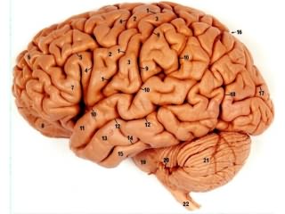
 Left side of the brain
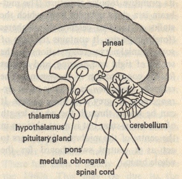 Mid cross section of the brain